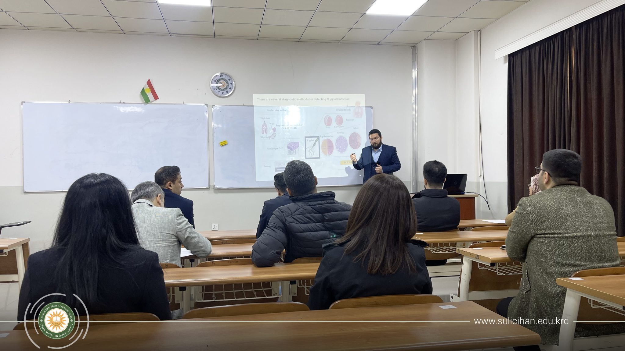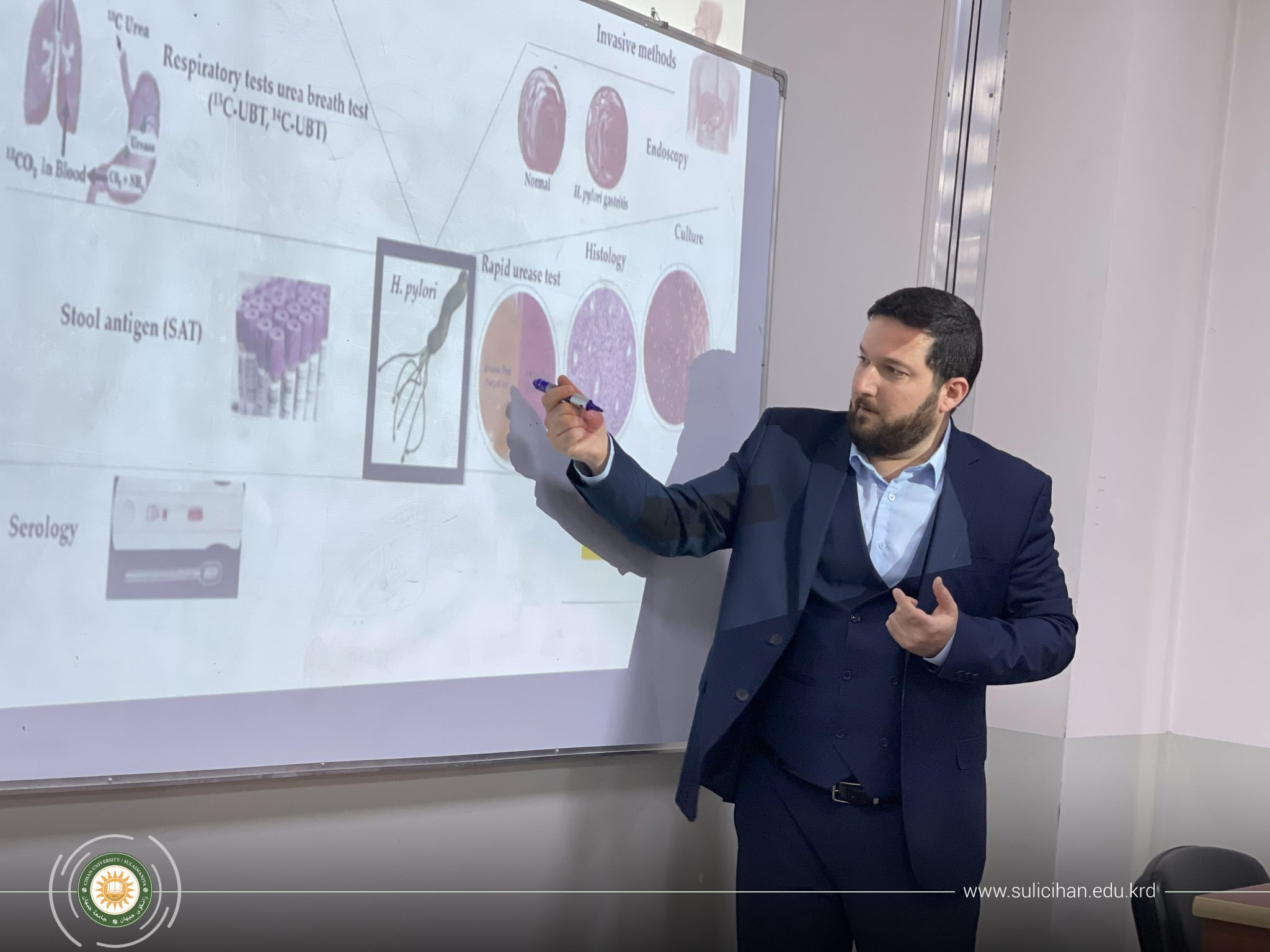
On Monday 12/23/2024, assistant lecturer Ahmed Ezzulddin Fakhruldin from the Department of Medical Microbiology at the College of Health Sciences presented a seminar about (Diagnostic Methods for Helicobacter pylori Infection).
The seminar discussed the methods used to detect Helicobacter pylori (H. pylori) Infection. H. pylori is a highly mobile spiral-shaped gram-negative bacterium with flagella at one pole. H. pylori grows in a microaerophilic atmosphere with low oxygen content and can easily survive in the stomach, despite the acidic environment. H. pylori can survive in the acidic environment of the stomach by producing urease, which catalyzes hydrolysis of urea to yield ammonia and CO2, the ammonia in turn elevates the pH around the bacteria in the stomach. H. pylori is considered the most common human pathogens and a leading etiological factor for various gastroduodenal diseases, including chronic gastritis, peptic ulcers, gastric adenocarcinoma, and mucosa-associated lymphoid tissue (MALT) lymphoma. Several methods are applied to detect H. pylori Infection which include invasive (endoscopy, histology, rapid urea test, and PCR), and non-invasive (Urea breath test, Stool antigen, and serology) methods. Moreover, the seminar discussed the advantages and disadvantages of invasive and non-invasive methods in the detection of H. pylori Infection.


